The Pintuition Marker
The power of a permanent magnet.
A rice-grain sized, non-radioactive marker in nickel-free biocompatible titanium. Its field never decays since it’s a permanent magnet, making the marker detectable from any direction, even in fluid, challenging lighting or near electrocautery. FDA-cleared for long-term placement, so the marker can be placed on the day of surgery or weeks to months in advance, allowing flexible scheduling.MDR CE marked for long-term placement, so the marker can be placed on the day of surgery or weeks to months in advance, allowing flexible scheduling.
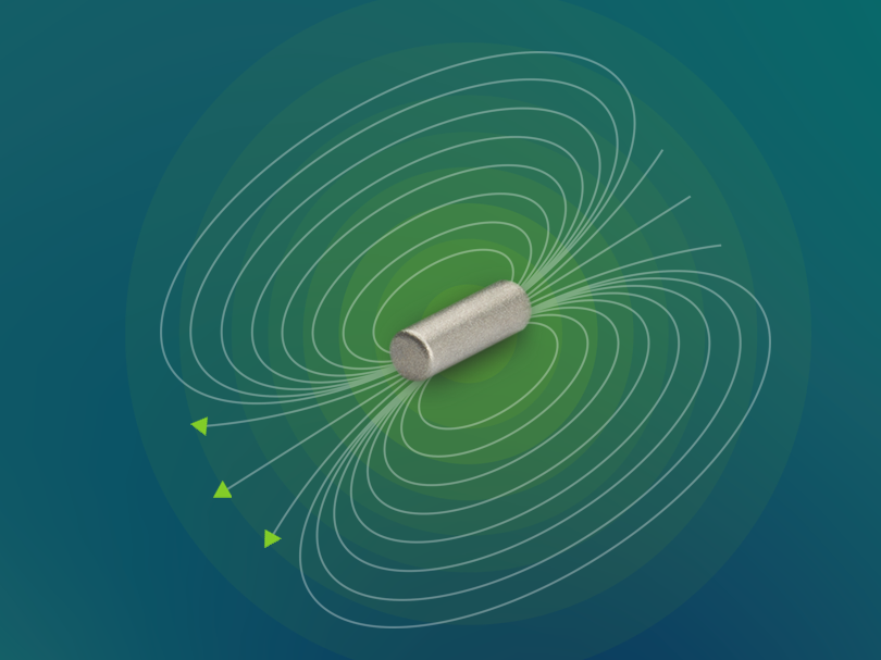
360° signal performance
Consistent signal detection regardless of marker orientation, providing reliable localization and accurate distance feedback.

No signal loss
A permanent magnet always has a field, even in fluids, challenging lighting or near electrocautery.

Robust marker
A high-grade biocompatible titanium housing ensures durability and reliable performance without nickel allergy concerns, whether placed on the day of surgery or months in advance.

Minimal discomfort
Intuitive, accurate devices that help clinicians deliver optimal care.

Non-radioactive
No nuclear medicine department or radiation-safety administrative burden.

- Marker5 × 1.6 mm, in a biocompatible titanium housing
- Needle14G, available in 7 and 12 cm lengths, with an ultrasound-enhanced tip
- IndicationAny soft-tissue localization including breast and lymph nodes; long-term implantation indicated for surgical removal
- VisibilityCompatible with all major imaging workflows, including ultrasound, X-ray (including stereotactic and contrast-enhanced mammography (CEM)), and CT
Advantages
Key advantages of using Pintuition as a Surgical Marker

Greater scheduling flexibility
Place the marker at any time — same day, or weeks to months before surgery.

Easy deployment
A 14G applicator in 7 and 12 cm lengths, preloaded with the marker and an ultrasound-enhanced tip.

Perfect visibility on imaging modalities
Compatible with all major imaging workflows, including ultrasound, X-ray (including stereotactic and contrast-enhanced mammography (CEM)), and CT.

Cost effective
Reduce delays, streamline workflows, and make better use of radiology and OR resources.
On imaging
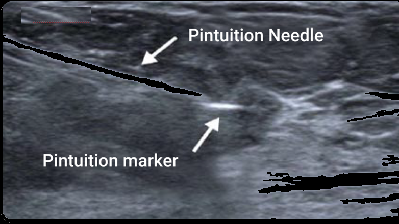
Pintuition Marker Visualization Across Imaging Modalities
Ultrasound
X-ray (mammography)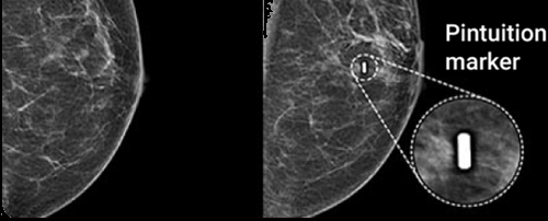
X-ray (specimen)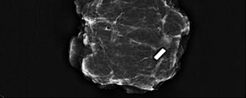
Contrast enhancement mammography (CEM)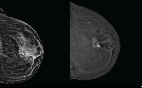
Axillary Lymph node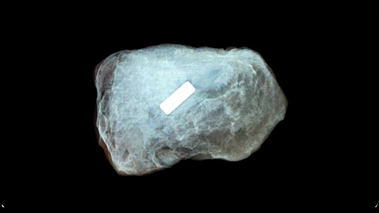
Trust and Embrace Surgical Marker Navigation
Radiologists and surgeons on Pintuition.


Dr. Floriaan Dijkman
Radiologist · ETZ Hospital, Netherlands
“We can now schedule up to a few weeks before the surgery, instead of placing the Pintuition marker on the day of the surgery or the day prior to surgery. So it gives more flexibility.”


Dr. Barbara Geeroms
Radiologist · UZ Leuven, Belgium
“The image visibility with ultrasound with the Pintuition Marker is very good. When you move the transducer a little, the marker is easily visible again because of its radiopacity. It’s also easily visible on the confirmation mammography.”


Dr. Suzanna van Veen
Radiologist · Rode Kruis Hospital, Netherlands
“The Pintuition marker placement can be planned one or two weeks in advance of surgery, which is not only convenient for the radiology department, but also for the patient.”


Matthew Miller, MD
Breast Surgeon · Valley Hospital Medical Center
“What really blew me away was the directionality. I had never worked with another localizer that could give me a three-dimensional direction to target.”


Shannon Tierney, MD, FACS
Breast Surgeon · Augusta Health Breast Surgery
“Because it’s extremely reliable and the targeting is so excellent, I know that my dissection is going in the correct direction from the get-go. I can choose to place my incision wherever I think it’s going to be the most cosmetic choice for the patient, and I don’t have to worry about compromising the oncologic resection.”


Renee Aboushi, MD
Breast Surgeon · Northwell Health
“What surprised me about using the Pintuition device was how easy it was to train surgical trainees on the localization tool. The Pintuition system fit into my surgical workflow seamlessly.”


Amelia Gunter, MD
Breast Surgeon · Medical City Weatherford
“We can place our incision where we want it and still find the marker and the tumor quickly.”
Dr. Floriaan Dijkman
Radiologist · ETZ Hospital, Netherlands
“We can now schedule up to a few weeks before the surgery, instead of placing the Pintuition marker on the day of the surgery or the day prior to surgery. So it gives more flexibility.”
Dr. Barbara Geeroms
Radiologist · UZ Leuven, Belgium
“The image visibility with ultrasound with the Pintuition Marker is very good. When you move the transducer a little, the marker is easily visible again because of its radiopacity. It’s also easily visible on the confirmation mammography.”
Dr. Suzanna van Veen
Radiologist · Rode Kruis Hospital, Netherlands
“The Pintuition marker placement can be planned one or two weeks in advance of surgery, which is not only convenient for the radiology department, but also for the patient.”


Dr. Janneke Berlage
Maasziekenhuis Pantein
“I only see advantages in it, you can operate more accurately which leads to better cosmetic outcomes.”


Dr. Louise de Widt
Dijklander Ziekenhuis
“With this new technique, the shortest path to the anomaly can be selected, and less tissue needs to be removed. This better preserves the breast, which is crucial for reconstruction.”


Miss Tamara Kiernan
Portsmouth Hospitals University NHS Trust
“It simply works every time.”

Dr. Gisela Moormann
Rode Kruis Ziekenhuis
“I think it’s a really great development. Of course for the patients, but the technical advantage for surgeons is also enormous.”
Get started with your Pintuition® trial
Try the Pintuition Marker in the clinic today and give your patients a better standard of breast cancer care with intuitive placement and minimal discomfort.
- With long-term placement indication the marker can be placed weeks or months ahead of surgery for complete scheduling flexibility.
- Compatible with all major imaging workflows, including ultrasound, X-ray and CT.
- No nuclear medicine department or radiation-safety administrative burden.
- 100% lesion localization & marker retrieval across 200 consecutive cases (Ceccato 2025).
- Approved for use in soft tissues, including the breast and axilla.
 Ordered through our partner Mammotome in the US & Canada.
80,000+Patients treated globally
Sirius Medical
Sirius Medical