Aug 21, 2026, 4:58:18 PM
Finding a firm, bruised swelling in your breast days after a biopsy is alarming, especially when the reason for the biopsy was a suspicious finding in the first place. In most cases that swelling is a hematoma, a collection of blood under the skin, and it heals on its own without treatment. This article explains how to recognize a hematoma after a breast biopsy, how long it takes to settle, what you can do at home, and which symptoms mean you should call your care team.
What Is a Hematoma After a Breast Biopsy?
A breast biopsy removes small samples of tissue through a needle, and like any procedure that passes through tissue it can nick small blood vessels along the way. When blood collects in the space left behind, it forms a hematoma: a firm, tender, often discolored swelling near the biopsy site. Breast tissue is well supplied with blood, which is why some bruising or a small hematoma is one of the most common side effects of the procedure. The American Cancer Society lists bruising and swelling among the expected effects of needle biopsies, and the larger the needle, the more likely some bleeding becomes. The different needle types are covered in Breast Biopsy Needle Techniques.
Bruise, Hematoma or Seroma: Telling Them Apart
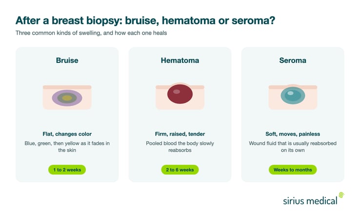
Three kinds of swelling can appear after a breast biopsy, and they behave differently. A bruise is flat discoloration of the skin that shifts from blue to green to yellow and fades within two weeks. A hematoma is a firm, raised, tender lump under the skin where blood has pooled, and it takes noticeably longer to resolve. A seroma is a soft, fluid filled pocket that is usually painless and moves slightly under the fingers. All three are common and usually harmless, but knowing which one you are feeling helps you describe it to your care team and saves unnecessary worry when a firm lump is still there after a month.
How Long Does a Breast Hematoma Take to Heal?
Most small hematomas soften and shrink over two to six weeks as the body gradually reabsorbs the blood. Larger ones can take a few months, and it is normal for the area to feel firm long after the discoloration has gone, because the body organizes the collected blood into denser tissue before clearing it. During that time the lump should slowly get smaller and less tender, not bigger and more painful. A hematoma that keeps growing in the first day or two after the biopsy is the exception and deserves prompt contact with your care team.
What Helps at Home
The practical advice is simple and makes a real difference in the first days:
- Apply a cold compress wrapped in a cloth for 15 to 20 minutes at a time during the first 24 to 48 hours to limit bleeding and swelling
- Wear a supportive, non wired bra day and night for the first days to keep pressure on the biopsy site
- Avoid strenuous exercise and heavy lifting for as long as your care team advises, typically a few days
- Use the pain relief your team recommends; check with them before taking medication that affects clotting, such as ibuprofen or aspirin, in the first days
- After the first two days, gentle warmth can help the body reabsorb the collected blood

Warning Signs: When to Call Your Care Team
- A swelling that keeps growing rapidly or becomes increasingly painful in the first 48 hours
- Increasing redness or warmth of the skin, fever above 38 degrees Celsius or 100.4 degrees Fahrenheit, or fluid leaking from the biopsy site, which can point to infection
- Skin that becomes tense, shiny or dark over the swelling
- A lump that has not clearly improved after six to eight weeks
None of these automatically mean something is wrong, but each is a reason to be seen rather than to wait.
Does a Hematoma Affect Your Biopsy Results or Marker?
The tissue samples were taken before the hematoma formed, so the swelling itself does not change your pathology results. What a hematoma can do is make follow up imaging harder to read: on ultrasound, a pocket of blood can obscure the area radiologists want to see. This matters because during most biopsies a small marker is placed at the sampled spot so it can be found again later, an approach explained in Clip Marker in Breast Cancer. If surgery follows, the surgeon needs to locate that exact area precisely, hematoma or not. This is one of the reasons detection technology matters: the magnetic detection used with the Pintuition Marker®, a permanent magnetic marker, keeps its accuracy in blood and fluid, so a hematoma around the biopsy site does not degrade the signal the surgeon navigates by. In the French 200 case series by Ceccato and colleagues in Scientific Reports (doi: 10.1038/s41598-025-88430-5), markers were placed days to weeks before surgery, 39.5 percent of them 2 to 29 days in advance, with 100 percent of lesions successfully excised. How markers are used to guide surgery is covered in Preoperative Localization in Breast Surgery.
Frequently Asked Questions
Is a hematoma after a breast biopsy dangerous?
Rarely. Most hematomas are self limiting and reabsorb without treatment. Very large or expanding hematomas occasionally need drainage, and signs of infection need review, but the typical firm bruise like lump after a biopsy heals by itself.
How long does bruising after a breast biopsy last?
Simple bruising fades within one to two weeks. A hematoma, which is a deeper collection of blood, usually needs two to six weeks and sometimes longer before the lump has fully gone.
Can a hematoma hide cancer on a mammogram?
A fresh hematoma can make imaging harder to interpret, which is one of the reasons radiologists place a marker during the biopsy and sometimes wait a few weeks before repeat imaging. The marker ensures the sampled area can always be identified regardless of the swelling. The UK spelling haematoma refers to the same finding.
Should a hard lump still be there a month after my biopsy?
A firm area at the biopsy site is common at one month and can persist while the body reorganizes and clears the collected blood. It should be getting slowly smaller and less tender. If it grows, hurts more, or simply worries you, contact your care team; a short ultrasound usually settles the question.
Can I fly or exercise with a breast hematoma?
Flying is generally not a problem once the first days have passed. For sport, follow your care team's advice: most people can resume light activity within days and strenuous exercise once the tenderness allows, usually within one to two weeks.
How Sirius Medical Supports Care After a Breast Biopsy
A biopsy is often the first step in a longer path, and the marker placed during that biopsy becomes decisive later, when a surgeon has to find and remove a lesion that cannot be felt. Sirius Medical developed the Pintuition System® for exactly that step. The Pintuition Marker® is a non radioactive, permanent magnetic marker that can be placed at the time of biopsy, and during surgery GPSDetect™ technology gives the surgeon continuous distance and direction to the marker, with a signal that stays accurate in blood, fluid and hematoma. That reliability in real world tissue conditions is part of why Elisabeth TweeSteden Hospital in the Netherlands has performed more than a thousand Pintuition® guided procedures. The broader marker landscape is described in Breast Markers in Breast Cancer.
Interested in Improving Surgical Localization?
Learn how Pintuition® can support precision, workflow efficiency and reliable excision in breast cancer surgery. Request demo or explore the clinical overview for more information.
Disclaimer
This article is intended for informational purposes only and should not be considered medical advice. Clinical decisions should always be made by qualified healthcare professionals based on individual patient needs and current guidelines.