

Nov 6, 2025 10:35:49 AM
Clip markers play an essential role in the diagnosis and surgical treatment of breast cancer. Placed during biopsy, these tiny markers ensure that suspicious lesions can be accurately located later during surgery or follow-up imaging. In this article, we explore what clip markers are, when and why they are used, and how they support procedures like targeted axillary dissection or breast-conserving surgery.
What Is a Clip Marker in Breast Cancer?
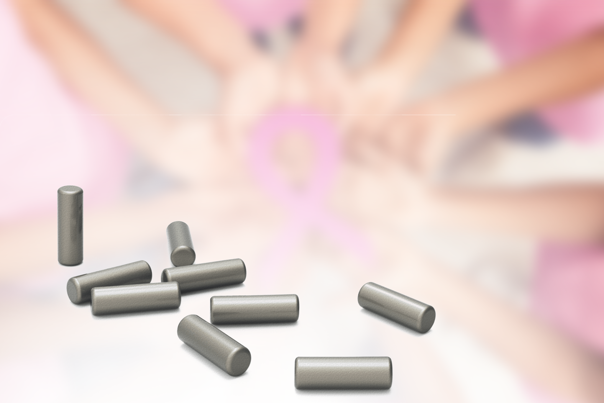
A clip marker is a small, biocompatible metallic or magnetic device placed in the breast at the time of biopsy. It remains in the tissue after sampling, allowing radiologists and surgeons to locate the original lesion even if it becomes invisible on imaging later. This is particularly important after neoadjuvant chemotherapy, where tumors may shrink or disappear radiologically.
Clip markers are commonly used in:
- Core needle biopsies or vacuum-assisted biopsies
- Multifocal or multicentric lesions requiring surgical planning
- Lesions expected to undergo response to systemic therapy
Learn more about biopsy devices and their clinical value.
Why Clip Marker Placement Matters
After biopsy, accurate placement of a clip marker allows for:
- Precise re-identification of the tumor bed at surgery
- Targeted axillary dissection in marked lymph nodes
- Improved coordination between radiology and surgical teams
- Use of intraoperative localization tools like magnetic seeds
Without a marker, it becomes difficult to identify the area of concern once the lesion responds to treatment. This can lead to larger excisions, more uncertainty, and higher re-excision rates.
What Happens After Marker Placement?
Once a clip marker is in place, it serves as the reference point for tumor localization. Before surgery, a localization procedure is performed to mark the clip’s location using a:
- Magnetic seed (e.g. Pintuition Marker®)
- Radioactive seed (RSL)
- Wire-guided localization
- Carbon injection (less common)
Read our full comparison: Seed vs Wire-Guided Localization.
Clip Markers and Lymph Node Surgery
Clip markers are also used to mark axillary lymph nodes that were proven metastatic during initial biopsy. After neoadjuvant therapy, these clips allow for re-identification of the same node, supporting procedures such as targeted axillary dissection (TAD).
This technique improves staging accuracy and helps avoid complete axillary lymph node dissection in eligible patients.
How Sirius Medical Supports Clip Marker Localization
While Sirius Medical does not provide clip markers, our localization technology directly supports the surgical phase following marker placement. The Pintuition Marker® is a magnetic seed that can be placed preoperatively at the site of the clip. During surgery, the Pintuition System® provides real-time directional feedback to guide accurate removal.
This enables:
- Safe and precise lesion resection
- Clear communication between radiologist and surgeon
- Flexible planning without radiation or same-day wire logistics
Pintuition® supports more than 35,000 breast cancer procedures worldwide.
Ready to improve your surgical localization workflow?
Request a demo or visit our resources page to download clinical guides and case studies.
Disclaimer
This article is intended for informational purposes only and should not be considered medical advice. While Sirius Medical is dedicated to improving breast cancer treatment through innovative localization technology, we do not provide medical diagnoses or treatment recommendations. If you experience any symptoms or changes in your breast health, consult a qualified healthcare professional promptly. Early medical evaluation is crucial for accurate diagnosis and effective treatment. Always seek professional guidance for concerns regarding your health.