Sep 21, 2026, 4:59:31 PM
Almost everyone who has a lumpectomy for breast cancer is advised to have radiation therapy afterwards, and the reason is straightforward: the operation removes the tumor, and radiation deals with what may be left behind in the rest of the breast. Together they achieve what a mastectomy achieves, while keeping the breast. This article explains why radiation follows breast conserving surgery, how long it takes, what the side effects really are, and one detail that is rarely explained: how the radiation oncologist knows exactly where the tumor used to be.
Why Radiation Follows a Lumpectomy
A lumpectomy, also called breast conserving surgery or wide local excision, removes the tumor with a rim of healthy tissue around it. The rest of the breast stays. Microscopic cancer cells can remain in that retained tissue, too few to see on imaging or to be caught by the pathologist, and radiation therapy is what deals with them.
The effect is well quantified. The Early Breast Cancer Trialists' Collaborative Group analyzed individual patient data from thousands of women in randomized trials and published the result in The Lancet (doi: 10.1016/S0140-6736(11)61629-2): radiotherapy after breast conserving surgery roughly halved the 10 year risk of any recurrence and reduced the 15 year risk of dying from breast cancer. That is why the combination of lumpectomy plus radiation, rather than lumpectomy alone, is the standard of care for early breast cancer. The comparison with removing the whole breast is set out in Lumpectomy vs Mastectomy.
A small number of women can safely skip radiation, typically older patients with small, hormone receptor positive tumors who will take endocrine therapy. That is a discussion to have with your oncologist rather than an assumption to make.
When Radiation Starts and How Long It Lasts
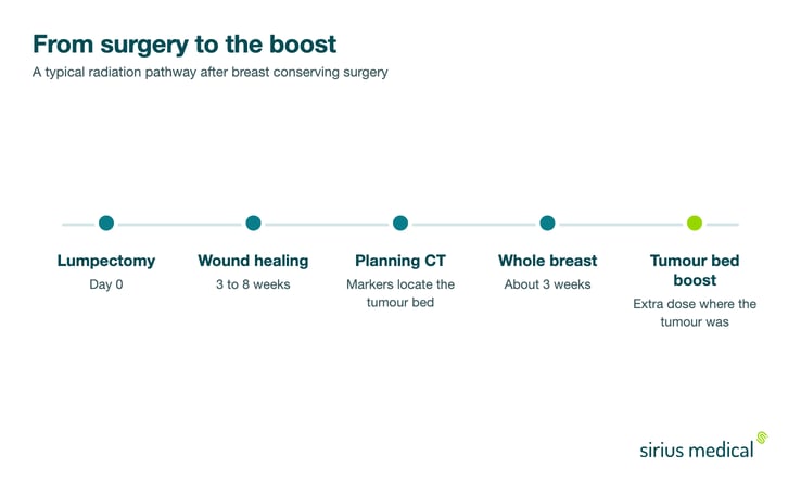
Radiation usually begins three to eight weeks after surgery, once the wound has healed. If chemotherapy is also planned, it normally comes first and radiation follows afterwards.
Schedules have shortened substantially. Where five to six weeks of daily treatment was once standard, most centers now use hypofractionated whole breast irradiation, which delivers a slightly higher dose per session over about three weeks. Some patients are eligible for partial breast irradiation, which treats only the area around the original tumor over an even shorter course. Sessions themselves are short, typically 10 to 20 minutes in the room with only a couple of minutes of actual beam time, and you go home straight afterwards. The American Cancer Society sets out the different schedules in more detail.
The Boost: Extra Dose Where the Tumor Was
Most local recurrences happen close to where the original tumor was, not scattered through the breast. That observation led to the boost: an additional dose aimed specifically at the tumor bed, the small volume of tissue the tumor was removed from.
The EORTC boost versus no boost trial, reported by Bartelink and colleagues in The Lancet Oncology (doi: 10.1016/S1470-2045(14)71156-8), followed patients for 20 years and found that the boost reduced local recurrence, with the largest benefit in younger women. It is now routine in many protocols.
A boost only works if the target can be defined accurately. After surgery the breast heals, tissue shifts and a seroma forms and then resolves, so the original tumor site is not obvious on a planning CT scan weeks later. Radiation oncologists therefore rely on markers: surgical clips placed in the walls of the cavity during the operation, or a marker already sitting at the lesion from the time of the biopsy. Those markers are what makes the boost field match the tissue actually at risk. The technique is covered in Tumor Bed Localization.
This is the part of the treatment chain that surprises people: a decision taken at the moment of biopsy, before anyone knows whether surgery will even be needed, still influences the accuracy of radiation planning months later. Why markers are placed at biopsy in the first place is explained in Clip Marker in Breast Cancer.
Side Effects, Short Term and Long Term
During treatment and in the first weeks afterwards, the common effects are:
- Skin redness, dryness, itching or a reaction resembling sunburn over the treated area, usually building from the second or third week
- Fatigue, which tends to accumulate over the course and can last several weeks after the final session
- Tenderness, aching or a heavy feeling in the breast
- Mild swelling of the breast
Longer term effects are less common and usually mild:
- Permanent slight darkening or thickening of the skin, and firmness of the breast tissue
- A small reduction in breast size or a change in shape
- Increased lymphedema risk when the armpit is included in the field, which is why the extent of nodal treatment matters; this is discussed in Lymphedema After Breast Cancer Surgery
- Rare effects on the underlying lung or, for left sided cancers, the heart. Modern planning techniques including breath hold have substantially reduced the dose reaching those organs
Questions Worth Asking Your Radiation Oncologist
- Which schedule am I having, and over how many weeks?
- Am I getting a boost to the tumor bed, and how is that area identified on my scan?
- Were surgical clips or a marker left at the site, and can they be seen on my planning scan?
- Will my lymph node areas be treated as well as the breast?
- For a left sided cancer, what is being done to limit the dose to my heart?
Frequently Asked Questions
Is radiation always needed after a lumpectomy?
Nearly always, because the combination of surgery and radiation is what makes breast conservation as safe as mastectomy. A limited group of older patients with small, hormone receptor positive tumors taking endocrine therapy may be able to omit it, after a specific discussion of the trade off.
How long after surgery does radiation start?
Typically three to eight weeks, once the wound has healed properly. If chemotherapy is part of the plan it usually comes first, which moves radiation several months further out.
Does radiation after lumpectomy hurt?
The treatment itself is painless; you feel nothing while the beam is on. Discomfort comes from the cumulative skin reaction and breast tenderness that build over the course, which is managed with skin care and settles in the weeks after the last session.
What is a tumor bed boost?
An extra dose delivered to the small area the tumor was removed from, because that is where most recurrences occur. It is given either as a few additional sessions after whole breast radiation or built into the main course, and it depends on markers or surgical clips to define the target accurately.
Can I have radiation if I had a marker or clip left in my breast?
Yes, and it usually helps. Markers and clips are designed to stay in place and are used by the radiation oncologist to identify the tumor bed on the planning scan. They do not interfere with the treatment.
What are the long term side effects of breast radiotherapy?
Most commonly mild changes in skin color and texture and some firmness of the breast tissue. Effects on the heart and lung are uncommon and have become less so with modern planning. Overall, serious late complications are rare relative to the reduction in recurrence risk.
Can I work during radiation treatment?
Many people do. Sessions are short and daily, so the main constraints are travel time and accumulating fatigue, which often becomes noticeable in the later weeks.
How Sirius Medical Contributes
Radiation planning is only as precise as the information left behind by the surgery and the biopsy that preceded it. Sirius Medical works at the start of that chain. The Pintuition Marker® is a non radioactive, permanent magnetic marker that can be placed at the time of biopsy and remain implanted long term, marking the lesion for the surgeon and leaving a durable reference point at the site.
During surgery the Pintuition System® uses GPSDetect™ to give the surgeon continuous distance and direction to the marker, with TargetLOC™ confirming alignment, so the excision is centered on the lesion rather than on an estimate of where it lies. In the French first 200 case series by Ceccato and colleagues in Scientific Reports (doi: 10.1038/s41598-025-88430-5), 100 percent of lesions were successfully excised and marker insertion accuracy was 100 percent. An accurately centered excision produces a cavity that genuinely corresponds to where the tumor was, which is what the radiation oncologist works from when planning the boost. How that site is defined for radiotherapy is described in Tumor Bed Localization, and the pathology side of the same question in Surgical Margins in Breast Conserving Surgery.
Interested in Improving Surgical Localization?
Learn how Pintuition® can support precision, workflow efficiency and reliable excision in breast cancer surgery. Request demo or explore the clinical overview for more information.
Disclaimer
This article is intended for informational purposes only and should not be considered medical advice. Clinical decisions should always be made by qualified healthcare professionals based on individual patient needs and current guidelines.