May 1, 2025 5:33:16 PM

When a breast abnormality is diagnosed, one of the next crucial steps is breast cancer localization. But how does this procedure work, and what are the different methods used for tumor localization?
What Is Breast Cancer Localization?
When a breast abnormality is diagnosed, breast cancer localization is often an essential next step in the treatment process. This procedure plays a critical role in guiding surgeons by precisely marking the area of concern before surgery. Breast cancer localization is commonly used in procedures such as lumpectomy or breast-conserving surgery, where the goal is to remove only the abnormal tissue while preserving as much healthy breast tissue as possible. The localization is always performed prior to surgery, with the timing depending on the specific method used.
During the procedure, a radiologist places a marker at the site of the abnormality. Then, during surgery, the surgeon uses this marker to accurately locate and remove both the marker and the abnormal tissue. Although the process may feel similar to a biopsy, its primary purpose is to place the marker, not to collect tissue samples.
Different Methods for Breast Cancer Localization
Breast cancer localization techniques generally fall into two main categories:
1. Wire Localization
Wire localization has been the standard method for surgical breast cancer localization since its introduction in the 1970s. This technique involves inserting a thin, flexible wire into the breast on the day of surgery. The wire serves as a precise guide for the surgeon, helping to accurately identify and remove the targeted area during the procedure. The external portion of the wire protrudes from the skin, allowing the surgeon to follow its path to the abnormal tissue.
Read more: Discover more about the disadvantages of wire-guided localization.
2. Wire-Free Localization
Wire-free localization involves placing a small marker directly into the abnormal tissue. Unlike wire localization, this method allows the procedure to be performed several days, or even up to 30 days, prior to surgery. During the operation, the surgeon uses a handheld device to detect the implanted marker and locate the targeted tissue with precision. This approach offers greater flexibility and improved patient comfort.
Several wire-free breast cancer localization techniques are used in clinical practice today to guide surgeons during breast-conserving procedures. These methods vary in terms of signal type, surgical workflow integration, and flexibility in timing. The choice of technique often depends on institutional protocols, equipment availability, and surgeon preference.
Radioactive Seed Localization
This method involves placing a very small seed that contains a low dose of radioactive material into the tumor. During surgery, a handheld probe detects the radiation emitted by the seed, helping the surgeon precisely locate and remove the abnormal tissue. Although effective, this method requires tight coordination between radiology, surgery, and nuclear medicine departments. Strict regulatory protocols must be followed to manage the safe storage, placement, and removal of the seed.
Infrared (Radar-Based) Localization
Infrared or radar-based systems use a small reflector implanted into the tumor. These reflectors are passive until activated during surgery by a probe that emits infrared or radar signals. The signal is reflected back to the probe, providing accurate information on the location of the tumor. These systems are non-radioactive, offering a safe and efficient alternative for surgical guidance.
Radio-Frequency Identification (RFID)
RFID-based localization involves inserting a small tag into the breast lesion prior to surgery. This is done by a radiologist under ultrasound or X-ray guidance. Each tag contains a unique identifier and emits a radio-frequency signal detectable by a handheld reader. This technique offers real-time detection and allows for placement days in advance, enhancing flexibility for both patients and surgical teams.
Magnetic Localization
Magnetic localization methods use a metallic marker that can be detected with a magnetic probe. There are two main types:
Some systems require the marker to be temporarily magnetized by an external probe just before detection. These rely on a form of iron detection and produce a signal only when the marker is magnetized.
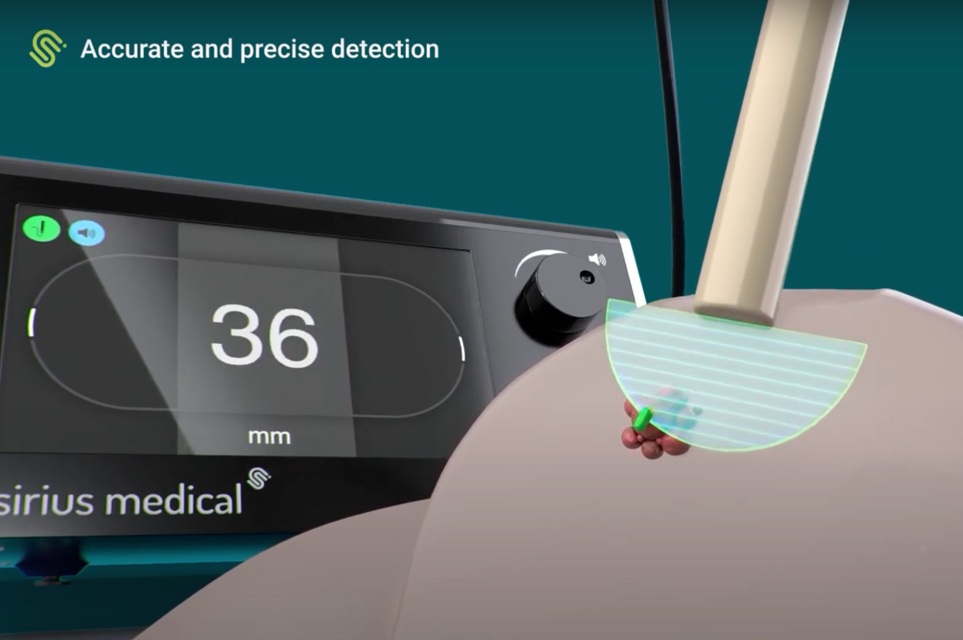
Other systems, such as permanent magnetic localization, use a marker that continuously emits a magnetic field. This allows the surgeon to locate the marker at any time without activation, improving consistency and simplifying the surgical workflow. The Pintuition® marker from Sirius Medical uses a permanent magnetic field to guide breast cancer localization. Unlike some other magnetic systems, the Pintuition marker does not require activation or magnetization: it continuously emits a magnetic signal, allowing for consistent and reliable detection.
Each wire-free localization technology offers specific advantages in terms of precision, patient comfort, and workflow integration. Factors such as surgical timing, regulatory requirements, and departmental coordination play a critical role in selecting the most suitable method.
Surgical Marker Navigation: Pintuition Marker®
The Sirius Pintuition® System marks a significant breakthrough in surgical technology, enhancing both precision and efficiency by replacing traditional breast cancer localization techniques with Surgical Marker Navigation for accurate tumor removal. At the core of this system is the Pintuition marker®: a small yet powerful magnetic marker that precisely marks the tumor's location, enabling surgeons to remove it with exceptional accuracy. Transitioning from wire-guided localization to the Pintuition system® streamlines collaboration and workflow between radiology and surgery departments. Powered by advanced navigation software, GPSDetect™, Sirius Pintuition® offers real-time directional guidance with both audio and visual cues, delivering unparalleled accuracy in tumor localization.

About Sirius Medical
With its roots deeply embedded in the Netherlands Cancer Institute, Sirius Medical is dedicated to improving care for cancer patients by delivering unsurpassed yet affordable solutions that enable precise and efficient removal of tumors. The Pintuition®technology is simple, precise, affordable, CE-marked, and FDA-cleared. Sirius Medical is rapidly expanding with over 35,000 procedures worldwide in more than 225 centers and a global commercial network covering the USA, Western Europe, Australia, and New Zealand.
Disclaimer
This article is intended for informational purposes only and should not be considered medical advice. While Sirius Medical is dedicated to improving breast cancer treatment through innovative localization technology, we do not provide medical diagnoses or treatment recommendations. If you experience any symptoms or changes in your breast health, consult a qualified healthcare professional promptly. Early medical evaluation is crucial for accurate diagnosis and effective treatment. Always seek professional guidance for concerns regarding your health.


