

Jan 27, 2026 9:14:41 PM
Breast markers are a foundational element of modern breast cancer care. They preserve the location of disease across diagnosis, treatment, and surgery, ensuring that lesions can be accurately identified even when imaging appearance changes. This article provides an in-depth overview of breast markers, how and when they are placed, and their clinical importance in contemporary breast cancer pathways.
What Are Breast Markers?
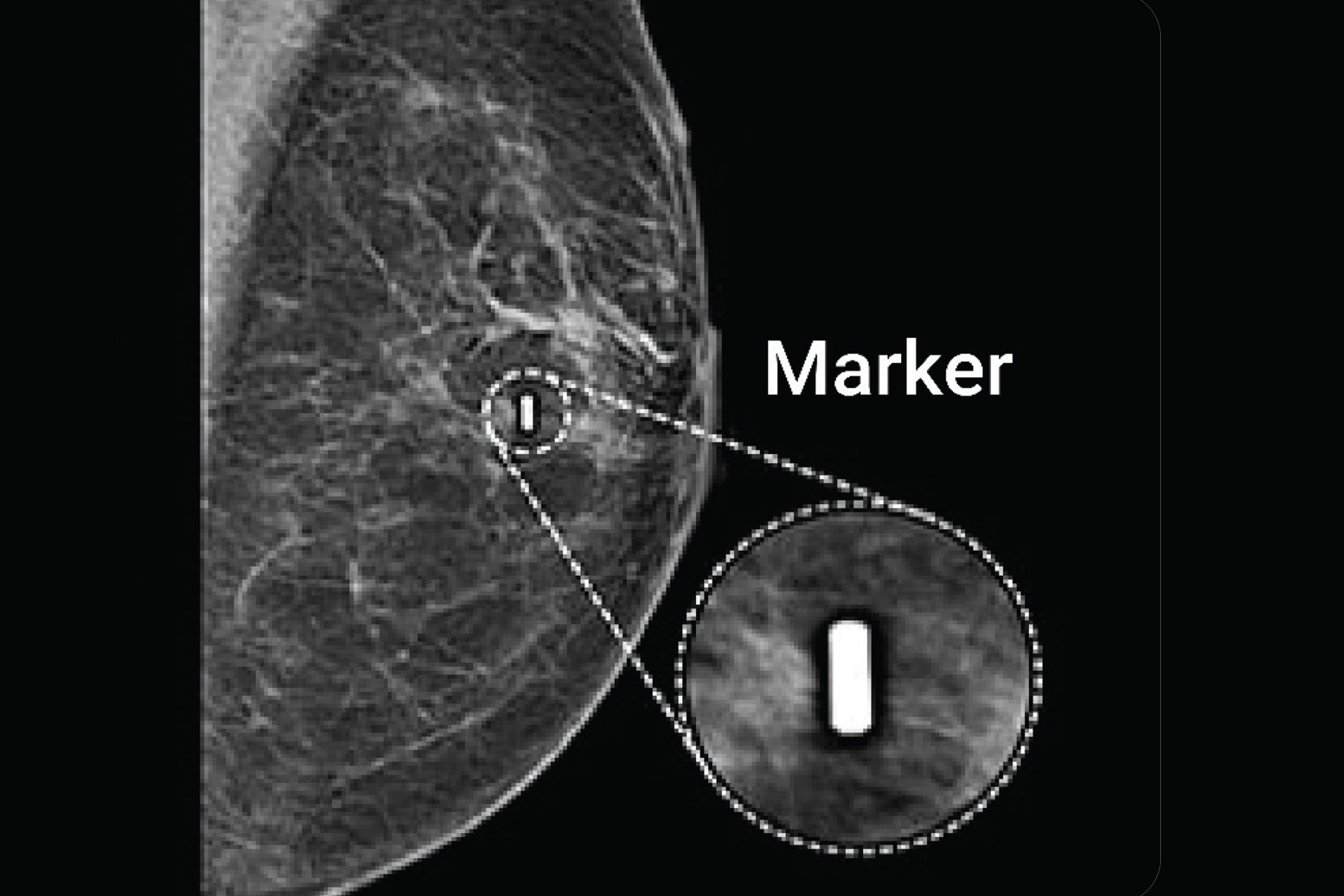
Breast markers are small medical devices placed in breast tissue or lymph nodes to mark the location of a lesion, biopsy site, or tumor bed. They are typically introduced during image-guided procedures and remain in place until surgery or long-term follow-up.
Markers are essential in cases where lesions are non-palpable, poorly visible on imaging, or expected to change due to neoadjuvant therapy. By preserving spatial information, breast markers enable continuity of care between radiology, pathology, and surgery.
Why Breast Markers Matter in Breast Cancer Care
Breast cancer treatment often spans weeks or months, during which the appearance of a tumor can change significantly. Without a marker, the original location of disease may become difficult or impossible to identify.
Breast markers support:
- Accurate correlation between diagnostic imaging and pathology
- Reliable localization of non-palpable tumors
- Preoperative planning for breast-conserving surgery
- Targeted resection after neoadjuvant systemic therapy
The clinical importance of localization is described in more detail in Breast Cancer Localization: Guiding Surgeons in the Removal of Abnormal Tissue.
Types of Breast Markers Used in Clinical Practice
Several categories of breast markers are used throughout the breast cancer pathway, each with a specific clinical role.
Biopsy Clip Markers
Clip markers are placed during core needle or vacuum-assisted biopsy to mark the exact site where tissue was sampled. They are especially important when the lesion is expected to shrink or disappear after treatment.
Clip visibility and placement considerations are discussed further in Breast Cancer Clip Visibility on Imaging.
Surgical Localization Markers
Before surgery, the biopsy clip often becomes the target for a localization procedure. Localization markers guide the surgeon to the correct area during the operation.
Traditional options include wire-guided localization, while newer approaches use non-radioactive or radioactive seeds. A clinical comparison is available in Seed Localization vs Wire-Guided: What Surgeons Need to Know.
Magnetic Seeds
Magnetic seeds are a newer category of breast markers designed to improve workflow and patient comfort. These small magnetic implants can be placed days or weeks before surgery and detected intraoperatively using a magnetic probe.
Magnetic seeds eliminate the need for same-day wire placement and avoid the regulatory burden associated with radioactive materials.
Placement of Breast Markers
Breast markers are placed under image guidance to ensure accurate positioning. The choice of imaging modality depends on lesion visibility and location.
- Ultrasound-guided placement for sonographically visible lesions
- Mammography or tomosynthesis-guided placement for calcifications
- MRI-guided placement for lesions visible only on contrast-enhanced imaging
More information on MRI-based workflows can be found in MRI-Guided Localization in Breast Cancer Surgery.
From Breast Markers to Surgical Localization
Markers alone do not complete the surgical pathway. They must be translated into actionable intraoperative guidance through localization.
This step bridges diagnostic imaging and surgical execution, enabling surgeons to confidently remove the intended tissue. An overview of localization strategies is available in Breast Cancer Localization.
Regulatory Advances and Expanded Clinical Use
In January 2026, Sirius Medical announced that its Pintuition® Surgical Marker Navigation System achieved MDR CE-mark certification. The Medical Device Regulation represents the most stringent regulatory framework for medical devices worldwide and confirms compliance with the highest safety and performance standards set by the European Commission.
As part of this certification, Pintuition® received expanded indications, including approval for long-term implantation. This enables radiology teams to place the Pintuition Marker® at the time of biopsy rather than immediately before surgery, offering greater flexibility in clinical planning.
Additional updates include expanded probe re-sterilization options and the availability of a shorter 7 cm needle, further broadening clinical applicability and improving operational efficiency.
How Sirius Medical Supports Breast Marker Localization
Sirius Medical focuses on translating marker placement into precise surgical guidance. The Pintuition Marker® is a non-radioactive magnetic seed designed to serve as a reliable reference point throughout the breast cancer care pathway.
During surgery, the Pintuition System®, enhanced by TargetLOC® technology, provides real-time distance and directional guidance, enabling surgeons to locate and remove non-palpable lesions with confidence.
This integrated approach supports accurate resection, predictable workflows, and improved coordination between radiology and surgery.
Interested in how breast markers fit into your surgical workflow?
Request a demo or explore our resources to learn how Sirius Medical supports precise breast cancer surgery.
Disclaimer
This article is intended for informational purposes only and should not be considered medical advice. Always consult a qualified healthcare professional.