

May 5, 2026 8:40:43 AM
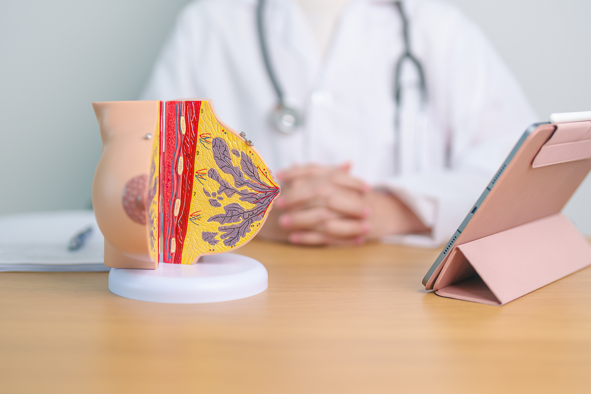
Image-guided surgery has transformed breast cancer treatment by allowing surgeons to operate on lesions that cannot be seen or felt directly. From the introduction of wire localization in the 1970s to the latest magnetic and electromagnetic navigation systems, image guidance has become integral to breast conserving surgery. This article explains what image-guided surgery means in a breast cancer context, how preoperative localization and intraoperative navigation work together, and how modern surgical marker navigation supports clear margins and predictable workflows.
What Is Image-Guided Surgery in Breast Cancer?
Image-guided surgery refers to surgical techniques that use imaging data to plan, locate and guide the resection of target tissue. In breast cancer, the technique is essential because most early stage tumors detected through screening are non palpable, meaning they cannot be located by touch alone. Without image guidance, the surgeon would have no reliable way to find the lesion, confirm its boundaries or limit the volume of healthy tissue removed.
For a foundational overview of how localization fits into breast cancer treatment, see Breast Cancer Localization.
Why Image Guidance Matters for Breast Cancer Surgery
Breast conserving surgery is the standard of care for most patients with early stage breast cancer, with survival outcomes equivalent to mastectomy when combined with adjuvant radiation. The clinical goal is to remove the tumor with clear margins while preserving as much healthy breast tissue as possible. Image-guided localization makes this possible for non palpable lesions and supports better oncologic and cosmetic results.
Research summarized on PubMed consistently shows that accurate preoperative localization improves margin outcomes and reduces the risk of re excision after breast conserving surgery.
The Two Phases of Image-Guided Breast Surgery
Image-guided breast surgery typically involves two phases. The first is preoperative localization, where a radiologist places a marker or guide near the lesion using ultrasound, mammographic or MRI guidance. The second is intraoperative navigation, where the surgeon uses a probe or system to find the marker during surgery and guide the resection.
Preoperative Localization
The radiologist selects an imaging modality based on lesion type, location and visibility. Ultrasound is often preferred for hypoechoic masses, mammography for calcifications and MRI for lesions only seen on MRI. For more on this topic, see MRI Guided Localization in Breast Cancer Surgery and Understanding Clip Marker Placement After Breast Biopsy.
Intraoperative Navigation
Once the patient is on the operating table, the surgeon uses guidance to locate and remove the targeted tissue. Older techniques rely on a wire that exits the skin, while newer systems use implantable markers detected by a handheld probe that provides distance or directional feedback in real time.
Localization Techniques in Image-Guided Surgery
Several localization techniques are currently used in clinical practice. Wire-guided localization remains widely available but requires same day placement, which limits scheduling flexibility. For a comparison, see Wire Guided Localization in Breast Surgery and Seed Localization vs Wire Guided.
Other techniques include radioactive seeds, magnetic markers, radar reflectors, radiofrequency tags and radio guided occult lesion localization (ROLL). Magnetic markers in particular have gained adoption because they are non radioactive, can be placed well before the day of surgery and integrate with simple intraoperative navigation. For background on this category, see Magnetic Seeds in Breast Cancer Surgery.
Workflow and Operating Room Efficiency
One of the practical advantages of modern image-guided systems is the ability to decouple radiology from surgery scheduling. With wire localization, both departments must coordinate on the same day, and a delay in one can disrupt the other. Markers that can be placed in advance allow each department to schedule independently, which improves operating room efficiency and reduces patient waiting time.
Guidance from organizations such as the American Cancer Society highlights the importance of coordinated multidisciplinary breast cancer care, where radiology, pathology and surgery work in alignment to support timely treatment.
Image-Guided Surgery After Neoadjuvant Therapy
Patients who receive neoadjuvant chemotherapy may experience significant tumor shrinkage, which can leave little or no imaging-detectable target at the time of surgery. In these cases, a marker placed before treatment becomes essential for identifying the tumor bed during breast conserving surgery. For more, see Tumor Bed Localization After Neoadjuvant Therapy.
How Sirius Medical Supports Image-Guided Breast Surgery
Sirius Medical developed the Pintuition System® to support image-guided breast cancer surgery from preoperative planning through intraoperative navigation. The Pintuition Marker® is a non radioactive magnetic marker placed under standard image guidance, with no restriction on the time interval between placement and surgery within the approved indication.
During surgery, the Pintuition System® provides surgeons with real time distance and directional feedback to the marker, supporting precise resection in both straightforward lumpectomies and more complex procedures including oncoplastic surgery. For a deeper look at our approach, see How Sirius Medical Oncology Medical Devices Support Breast Cancer Care.
Interested in Improving Surgical Localization?
Learn how Pintuition® can support precision, workflow efficiency and confidence in image-guided breast cancer surgery. Request a demo or explore the clinical overview for more information.
Disclaimer
This article is intended for informational purposes only and should not be considered medical advice. Clinical decisions should always be made by qualified healthcare professionals based on individual patient needs and current guidelines.