

Jun 16, 2026 8:33:13 AM

The success of breast conserving surgery is judged largely on one pathology result: the margin. A clear margin means the surgeon removed the cancer with a rim of healthy tissue around it. A positive margin means cancer cells reach the cut edge, and it usually sends the patient back for a second operation. Margins sit at the intersection of oncology, cosmetics and hospital logistics, and the way a lesion is localized has a direct and now well documented effect on the outcome. This article explains what a clear margin actually is, why a positive one costs far more than a second surgery, and how localization quality changes the odds of getting it right the first time.
What Surgeons Actually Mean by a Clear Margin
There is a common misconception that a wider margin is always better. Current practice says otherwise. For invasive breast cancer, the widely adopted consensus standard is no ink on tumor, meaning the cancer does not touch the inked outer surface of the removed specimen. For ductal carcinoma in situ, the standard is wider, typically at least 2 mm of clear tissue.
The reason the two standards differ is biological. Invasive cancer that has been fully excised with adjuvant radiation does not benefit from removing extra healthy tissue beyond a clear edge, so larger margins add cosmetic harm without oncologic gain. In situ disease spreads along the duct system in a less predictable, sometimes discontinuous pattern, which is why a measurable buffer is recommended. For more on the in situ scenario, see DCIS Surgery and Localization. Guidance reflecting these standards is summarized by the National Comprehensive Cancer Network.
The Real Cost of a Positive Margin
A positive margin is often described simply as a reason for a second surgery, but the true cost is broader. A re excision means a second exposure to anesthesia, additional recovery, and a delay before adjuvant therapy such as radiation can begin. It compounds patient anxiety at an already difficult moment. It often produces a worse cosmetic result, because a second resection in the same area removes more tissue and disturbs healing. And at the level of the hospital it consumes a second operating slot, imaging, and pathology, raising the cost of treating that patient considerably.
Seen this way, the margin result is not just a surgical detail. It is the single decision point that determines whether a breast conserving pathway stays on track or doubles in length and cost. Anything that reliably increases first attempt clearance has value across all of those dimensions.
Why Localization Technique Influences Margin Outcomes
It is worth being precise about the mechanism, because the marker itself does not cut the tissue. Localization affects margins in two ways. Before surgery, an accurately placed marker lets the surgeon plan the resection in three dimensions around the real target rather than around an approximate area. During surgery, a system that reports distance and direction to that marker lets the surgeon judge how much tissue lies between the target and the planned cut surface, in real time, rather than estimating by feel.
Traditional approaches limit one or both of these. A wire indicates a path but gives limited directional information once the operation begins, and it must be placed on the day of surgery. For a comparison of approaches, see Wire Guided Localization in Breast Surgery and Seed Localization vs Wire Guided.
What the Evidence Shows
Two recent peer reviewed studies make the margin case concretely. The first 200 case French series by Ceccato and colleagues in Scientific Reports (doi: 10.1038/s41598-025-88430-5) reported a re excision rate of 9 percent with magnetic surgical marker navigation, against a pooled wire guided benchmark of 14.9 to 20.8 percent, and comparable to radioactive seed benchmarks. A comparative study by Chinn and colleagues at Stanford University in Annals of Surgical Oncology (doi: 10.1245/s10434-025-18354-x) reported a positive margin rate and re excision rate of just 2.2 percent.
Clear Margins Without Removing More Tissue
The most instructive finding is one that is easy to overlook. In the Stanford comparison, the volume of tissue removed was statistically similar between the two technologies studied (around 32 cubic centimeters versus 27, with a p value of 0.80), yet margins and re excision rates were identical and operative time was shorter with Pintuition®. This matters because it shows that better margin performance does not have to come from simply cutting out more breast. The goal in breast conservation is the opposite: clear margins with maximum tissue preservation. Accurate navigation supports both at once rather than trading one for the other. A study summary is available in the clinical one pager.
The Probe as an Intraoperative Ruler
One practical detail from the French series captures how navigation supports margins in the moment. Surgeons reported using the probe to check the distance between the resection margin and the marker before completing the excision. Rather than waiting for pathology to discover a close margin after the fact, the surgeon gains an intraoperative estimate of how much tissue sits between the target and the edge. For research context on margin assessment, see literature indexed on PubMed.
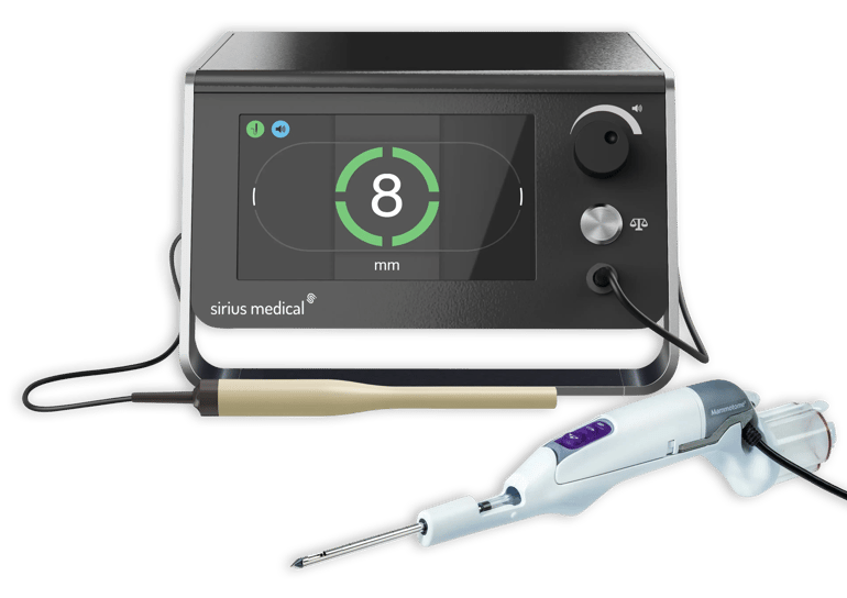
How Sirius Medical Supports Clear Margins
Sirius Medical developed the Pintuition System® to give surgeons real time, bi directional distance and directional feedback to the Pintuition Marker® with millimeter level accuracy. The marker is non radioactive and placed under standard image guidance, and the signal remains accurate in the presence of blood or fluid and is unaffected by electrocautery, so navigation continues to work in exactly the conditions where margin decisions are made. For the wider clinical picture, see How Sirius Medical Oncology Medical Devices Support Breast Cancer Care.
Interested in Improving Surgical Localization?
Learn how Pintuition® can support clear margins, tissue preservation and first attempt success in breast conserving surgery. Request a demo or explore the clinical overview for more information.
Disclaimer
This article is intended for informational purposes only and should not be considered medical advice. Clinical decisions should always be made by qualified healthcare professionals based on individual patient needs and current guidelines.